Opera Phenix® Plus is a high throughput microplate confocal imager for High-Content Screening (HCS).

It can acquire, analyze and manage fluorescence, brightfield and digital phase contrast images.

⚙️ Configuration

📷 Two sCMOS Cameras

  • 7 megapixels (2160×2160), 16-bit resolution, 6.5 μm pixel size
  • Simultaneous acquisition of up to 4 channels with minimized crosstalk
  • Each camera supports fast recording mode: up to 100 frames per second (fps)

20220718134245.135.png


🔍 Synchrony Optics combines

  • Microlens-enhanced Nipkow spinning disk with
  • Dual-view confocal optics to separate fluorescence excitation and emission during simultaneous acquisition

💡 Five excitation Lasers

  • 375 nm / 50 mW
  • 425 nm / 100 mW
  • 488 nm / 150 mW
  • 561 nm / 100 mW
  • 640 nm / 140 mW

🖼️ Imaging Modalities

  • Switchable widefield and confocal imaging, based on assay requirements
  • Micro lens-enhanced dual spinning disk with optimized pinhole spacing for reduced background in thick samples (e.g., spheroids)

🌕 Transmission Brightfield & Digital Phase Contrast

🎯 Emission Filter

  • 435–515 nm, typically used for CFP/YFP FRET studies

🔬 Objectives

Air Objectives:

  • 5×, NA 0.16, WD 12.1 mm
  • 10×, NA 0.3, WD 5.2 mm
  • 20×, NA 0.4, WD 8.28 mm

💧 Water Immersion Objectives provides an improved light collection efficiency:

  • 20×, NA 1.0, WD 1.7 mm
  • 40×, NA 1.1, WD 0.62 mm
  • 63×, NA 1.15, WD 0.6 mm


🌡️ Live Cell Chamber

  • Controlled temperature, CO₂, and humidity – ideal for live cell experiments

🧪 Plate Compatibility

  • Compatible with various well plates such as 24, 96- and 384-well plates
  • Slide holder: up to 4 × 4 slides

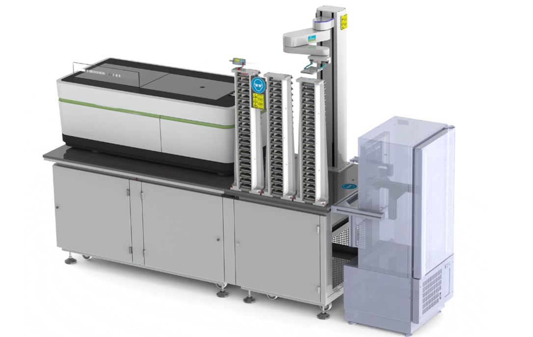
Live Cell Imaging Package

  • FLEX 750 collaborative robotic plate handler
  • Control PC, barcode reader, and PlateWorks™ scheduling software
  • 14-position plate shelf
  • Liconic STX44 Incubator
    • Temperature range: 32–50 °C
    • Humidity >95%, CO₂ 0–10%
    • Two 22-position plate cassettes for 96/384-well plates

20220718134353.739.png

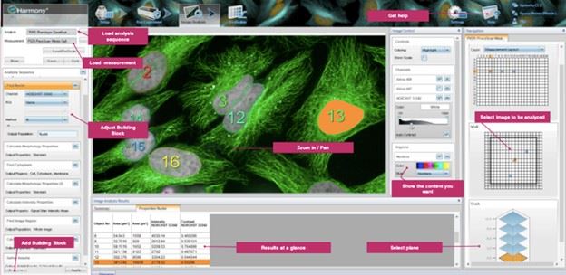
🧠 Harmony® High-Content Imaging and Analysis Software

✨ Advanced analysis features such as texture analysis and STAR morphology provide detailed characterization of cellular phenotypes, enabling more robust and reproducible classification

🧩 Powerful 3D visualization and analysis features

🧱 Ready-made solutions and modular building blocks for image analysis workflows

🤖 Includes 2× PhenoLOGIC™ – machine learning algorithm –based phenotype classification

🎯 Includes 2× PreciScan™ – intelligent acquisition plug-in that identifies the object's xyz-position and enables targeted re-scanning

🧬 Includes support for Cell Painting assays

20250622134825262.jpg